
Brain anatomy is the study of the brain’s structure and organization, essential for understanding its functions and disorders. It reveals how the brain controls motor, sensory, and cognitive processes, making it vital for medical and scientific research.
Resources like brain anatomy PDF guides provide detailed visuals and explanations, aiding students and professionals in mastering this complex field.
Understanding brain anatomy is crucial for diagnosing and treating neurological conditions, as well as advancing neuroscience research and education.
1.1 Overview of Brain Structure
The brain’s structure is highly organized, consisting of three main regions: the cerebrum, cerebellum, and brainstem. The cerebrum, the largest part, manages sensory and cognitive functions, while the cerebellum coordinates movement. The brainstem connects the brain to the spinal cord, regulating vital functions like breathing. Brain anatomy PDF guides often highlight these divisions, providing detailed diagrams and explanations. Understanding this structural framework is essential for grasping how the brain operates and how disorders develop. These resources are invaluable for both educational and clinical purposes, offering a clear visual and textual breakdown of brain anatomy.
1.2 Importance of Understanding Brain Anatomy
Understanding brain anatomy is crucial for advancing neuroscience, improving diagnostics, and developing treatments for neurological disorders. It helps explain how the brain controls functions like movement, cognition, and emotion. Brain anatomy PDF guides are invaluable for education and research, offering detailed insights into structural components. This knowledge aids in identifying abnormalities and understanding disease mechanisms, ultimately enhancing patient care and therapeutic outcomes. By studying brain anatomy, professionals can better address conditions such as stroke, tumors, and neurodegenerative diseases, making it a cornerstone of medical and scientific progress.
Major Regions of the Brain
The brain is divided into three main regions: the cerebrum, cerebellum, and brainstem. Each plays a vital role in controlling different bodily functions and processes.
2.1 Cerebrum
The cerebrum is the largest part of the brain, divided into two hemispheres. It controls voluntary movements, processes sensory information, and manages higher cognitive functions like memory and decision-making.
It is further divided into four lobes: frontal, parietal, temporal, and occipital, each specializing in different functions.
Damage to the cerebrum can affect motor skills, speech, and mental abilities, highlighting its critical role in human consciousness and behavior.
2.2 Cerebellum
The cerebellum is located at the back of the brain and plays a key role in motor coordination, balance, and posture. It ensures smooth and precise movements by refining muscle activity.
Damage to the cerebellum can lead to ataxia, characterized by unsteady movements and loss of coordination. The cerebellum also contributes to learning and memory, particularly in motor skills. Its structure includes the cerebellar cortex and deep nuclei, which process sensory and motor information. Understanding its functions is crucial for diagnosing and treating cerebellar-related disorders, as detailed in brain anatomy PDF resources.
2.3 Brainstem
The brainstem connects the cerebrum to the spinal cord, acting as a vital relay center for sensory and motor signals. It consists of the midbrain, pons, and medulla oblongata.
The brainstem regulates essential functions such as breathing, heart rate, and blood pressure. It also manages alertness, arousal, and the sleep-wake cycle. Damage to the brainstem can result in severe impairments or even death. Brain anatomy PDF guides often highlight its critical role in maintaining basic life-sustaining activities, making it a fundamental area of study in neuroscience and neurology.
Subdivisions of the Cerebrum
The cerebrum is divided into two hemispheres and four lobes: frontal, parietal, temporal, and occipital. These subdivisions specialize in motor, sensory, and cognitive functions. Brain anatomy PDF guides detail these structures, aiding in comprehensive understanding and visualization of their roles in brain organization and function.
3.1 Lobes of the Brain
The brain is divided into four lobes: frontal, parietal, temporal, and occipital. Each lobe specializes in specific functions, such as decision-making, sensory processing, memory, and vision.
Brain anatomy PDF guides provide detailed diagrams and descriptions of these lobes, helping to visualize their roles and interconnectedness.
Understanding the lobes is crucial for comprehending brain function, as they collectively enable motor skills, sensory perception, and cognitive processes.
3.2 Cortical Layers
The cerebral cortex is organized into distinct layers, typically six, each containing specific types of neurons and neural connections. These layers play a critical role in processing sensory information, controlling movement, and enabling complex thought.
Brain anatomy PDF guides often include detailed cross-sectional views of cortical layers, helping to visualize their arrangement and function.
Understanding cortical layers is essential for mapping brain function to structure, aiding in the study of neurological disorders and brain injuries.
3;3 White and Gray Matter
The brain’s structure comprises gray matter and white matter, each fulfilling distinct roles. Gray matter contains neurons that process information, while white matter consists of myelinated axons facilitating communication between brain regions.
Brain anatomy PDF guides provide detailed visuals of these structures, highlighting their roles in sensory and motor functions;
Understanding the differences between gray and white matter is vitally essential for studying brain development and neurological disorders.

Functions of the Brain
The brain controls motor, sensory, and cognitive processes, enabling thought, movement, and emotional regulation. It integrates information to maintain bodily functions and facilitate complex behaviors, as detailed in brain anatomy PDF guides.
4.1 Sensory and Motor Functions
The brain’s sensory functions process information from sensory organs, interpreting stimuli like touch, vision, hearing, taste, and smell. Motor functions involve controlling voluntary movements, such as walking or writing, through neural signals transmitted to muscles and glands. Brain anatomy PDF guides highlight how different brain regions, like the cerebral cortex and brainstem, coordinate these functions. Damage to these areas can lead to sensory deficits or motor impairments, emphasizing the brain’s critical role in maintaining these essential bodily operations.
Understanding these processes is vital for diagnosing and treating neurological disorders.
4.2 Cognitive Functions
Cognitive functions encompass mental processes like memory, attention, problem-solving, and language. The prefrontal cortex manages decision-making, while the temporal lobes play a key role in memory formation. Brain anatomy PDF guides illustrate how these regions interact to enable learning and reasoning. Damage to these areas can impair cognitive abilities, highlighting the brain’s intricate organization. Understanding these functions is essential for addressing conditions like dementia and developing strategies to enhance cognitive performance.
These processes are fundamental to human thought and behavior, making them a cornerstone of brain anatomy studies.
4.3 Emotional Regulation
Emotional regulation is managed by brain structures like the amygdala, prefrontal cortex, and limbic system. The amygdala processes emotions, while the prefrontal cortex modulates emotional responses. Brain anatomy PDF guides detail these regions, showing how they interact to balance emotional states. Dysregulation in these areas can lead to disorders like anxiety or depression. Understanding this anatomy is vital for addressing mental health conditions and developing therapeutic strategies to manage emotions effectively.
Emotional regulation is a complex process that underscores the brain’s role in maintaining psychological well-being.
Blood Supply to the Brain
The brain receives blood through two main arterial systems: the carotid and vertebral arteries. These supply oxygen and nutrients, ensuring proper brain function and survival.
5.1 Arterial Supply
The brain’s arterial supply primarily comes from the carotid and vertebral arteries. The carotid arteries divide into internal and external branches, with the internal carotid supplying the anterior brain. The vertebral arteries merge to form the basilar artery, which supplies the posterior brain. These arteries converge at the circle of Willis, a critical structure near the base of the brain, ensuring a rich blood supply to cerebral tissues. This network is vital for delivering oxygen and nutrients, maintaining brain function and survival.
5.2 Venous Drainage
Venous drainage of the brain primarily occurs through the dural venous sinuses, which are located within the dura mater. These sinuses collect deoxygenated blood from cerebral veins and direct it toward the internal jugular veins. The superior sagittal sinus, the largest dural sinus, runs along the midline of the brain and drains into the confluence of sinuses near the occipital bone. Proper venous drainage is essential for maintaining brain health, as it removes waste products and prevents cerebral swelling. This system ensures efficient blood circulation and supports overall neurological function;
Neuroimaging Techniques
Neuroimaging techniques provide detailed visualization of brain structures and functions, aiding in both research and clinical diagnostics by capturing high-resolution images of the brain’s anatomy and activity.
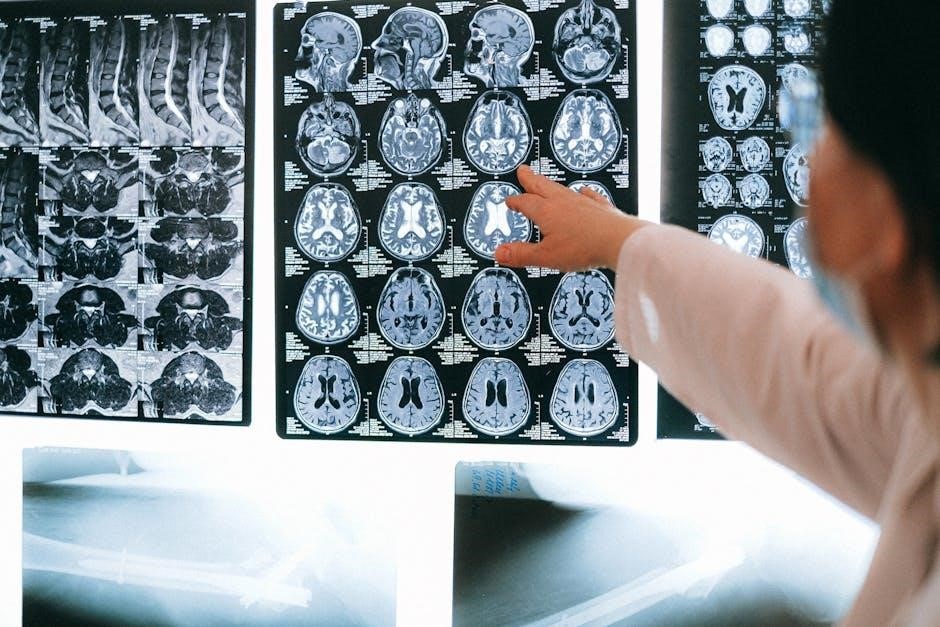
6.1 MRI and CT Scans
Magnetic Resonance Imaging (MRI) and Computed Tomography (CT) scans are widely used neuroimaging techniques that provide detailed images of brain structures. MRI uses magnetic fields and radio waves to generate high-resolution images of soft tissues, including gray and white matter, while CT scans employ X-rays to produce cross-sectional images of the brain, particularly useful for detecting bone abnormalities and acute injuries. Both methods are non-invasive and complement each other in diagnosing brain disorders, making them essential tools in clinical and research settings for understanding brain anatomy and pathology.
6.2 PET and SPECT Scans
Positron Emission Tomography (PET) and Single Photon Emission Computed Tomography (SPECT) are functional neuroimaging techniques that visualize brain activity and metabolism. PET scans use radioactive tracers to map glucose uptake, oxygen consumption, or neurotransmitter activity, aiding in diagnosing Alzheimer’s disease, epilepsy, and brain tumors. SPECT scans also employ tracers to assess blood flow, commonly used in stroke evaluation and psychiatric disorders. Both methods provide complementary data to structural imaging like MRI and CT, offering insights into brain function and pathology, enhancing diagnostic accuracy and treatment planning in clinical and research settings.
Brain Disorders and Diseases
Brain disorders include structural, neurodegenerative, and psychiatric conditions. Examples are Alzheimer’s disease, stroke, and schizophrenia. Understanding brain anatomy aids in diagnosing and treating these conditions effectively.
7.1 Structural Disorders
Structural brain disorders involve abnormalities in brain tissue or architecture, often due to injury, infection, or congenital conditions. Examples include brain tumors, hydrocephalus, and cerebral edema. These disorders can disrupt normal brain function, leading to symptoms like seizures, cognitive decline, or motor impairments. Understanding brain anatomy through resources like brain anatomy PDF guides helps identify and classify structural abnormalities. Diagnosis often relies on imaging techniques such as MRI or CT scans, which visualize structural changes. Early detection and treatment are crucial for managing these conditions and improving patient outcomes.
7.2 Neurodegenerative Diseases
Neurodegenerative diseases are progressive conditions characterized by the loss of brain cells and tissue, leading to cognitive, motor, and functional decline. Alzheimer’s disease, Parkinson’s disease, and Huntington’s disease are prominent examples. These disorders often result from protein misfolding or genetic mutations, causing irreversible damage to brain structures. Brain anatomy resources, such as detailed brain anatomy PDF guides, help visualize affected areas and understand disease progression. Early diagnosis through neuroimaging and clinical evaluation is critical for managing symptoms and improving quality of life, though treatments remain largely symptomatic.
7.3 Psychiatric Disorders
Psychiatric disorders, such as schizophrenia, depression, and anxiety, are linked to abnormalities in brain structure and function. Brain anatomy studies reveal altered volumes in regions like the prefrontal cortex and amygdala, which regulate emotions and decision-making. These changes, often visualized through neuroimaging, help clinicians understand the biological basis of mental health conditions. Brain anatomy PDF guides provide detailed insights into these structural variations, aiding in diagnosis, treatment planning, and research into psychiatric illnesses. Understanding brain anatomy is crucial for developing targeted therapies and improving outcomes for individuals with mental health disorders.

Comparative Brain Anatomy
Comparative brain anatomy explores differences and similarities between human and animal brains, offering insights into evolutionary development and functional adaptations. Brain anatomy PDF guides highlight these comparisons, aiding researchers in understanding cognitive and structural variations across species.
8.1 Human vs. Animal Brains
Human brains are larger and more complex compared to animal brains, with advanced cognitive functions like language and abstract thinking. Brain anatomy PDF guides reveal that humans have a higher brain-to-body mass ratio and a more developed cerebral cortex, enabling superior problem-solving abilities. In contrast, animal brains prioritize instinctual behaviors and sensory processing. While both share basic structures like the brainstem and cerebellum, humans possess unique features such as enlarged prefrontal lobes. These differences highlight evolutionary adaptations, making human brain anatomy distinct yet fundamentally connected to other species.
8.2 Evolutionary Perspectives
Brain anatomy has evolved significantly over millions of years, with humans developing larger, more complex brains compared to other species. The cerebral cortex expanded, enabling advanced cognitive functions like language and abstract thought. Encephalization, or increased brain-to-body mass ratio, drove human adaptation and survival. Brain anatomy PDF guides highlight how evolutionary pressures shaped structures like the prefrontal cortex for decision-making and innovation. Studying these developmental pathways provides insights into how human cognition diverged from other animals, emphasizing the brain’s role in human dominance and cultural advancement.

Clinical Relevance of Brain Anatomy
Understanding brain anatomy is crucial for neurosurgeons and neurologists, aiding in precise brain mapping and diagnosis. Brain anatomy PDFs serve as essential tools for clinical visualization and education.
9.1 Neurosurgery
Brain anatomy PDFs are essential for neurosurgeons, providing detailed maps of brain structures. These resources aid in pre-surgical planning and intraoperative navigation, ensuring precision in complex procedures.
They help identify functional areas, such as motor and language centers, minimizing risks to critical brain regions.
Additionally, brain anatomy PDFs are used to educate patients and train future surgeons, enhancing understanding of neurosurgical approaches and techniques.
9.2 Neurology
Brain anatomy PDFs are invaluable in neurology for diagnosing and managing neurological disorders. They provide detailed visualizations of brain structures, aiding in identifying abnormalities such as tumors, strokes, or degenerative changes.
These resources help neurologists correlate structural anomalies with clinical symptoms, enhancing diagnostic accuracy.
Additionally, brain anatomy PDFs are used to educate patients about their conditions and treatments, fostering better understanding and engagement in care. They also serve as essential tools for neurology residents and practitioners to refine their knowledge of brain organization and function.

Educational Resources for Brain Anatomy
Brain anatomy PDFs are comprehensive guides offering detailed diagrams and explanations of brain structures. They are widely used in classrooms and self-study, making complex concepts accessible and engaging for learners. These resources often include labeled illustrations, functional overviews, and clinical correlations, catering to students, educators, and professionals. Digital accessibility ensures easy reference, enhancing the learning experience and fostering a deeper understanding of brain anatomy. They are indispensable tools for both academic and professional development in neuroscience and related fields.
10.1 Brain Anatomy PDF Guides
Brain anatomy PDF guides provide detailed, visually rich resources for understanding brain structure. They include labeled diagrams, cross-sectional views, and functional explanations. These guides are ideal for students, researchers, and professionals, offering a portable and accessible way to study brain anatomy. Many PDFs are available online, catering to various learning levels, from introductory to advanced. They often cover topics like cortical layers, brain regions, and neuroanatomical landmarks. These resources are invaluable for education, research, and clinical reference, making complex brain anatomy concepts more engaging and easier to comprehend.
10.2 Online Courses and Tutorials
Online courses and tutorials offer interactive and comprehensive learning experiences for brain anatomy. Platforms provide structured lessons, quizzes, and multimedia content, making complex concepts engaging. These resources cater to diverse learning levels, from beginners to advanced learners. Many courses include 3D models, video lectures, and downloadable materials, enhancing understanding. They are particularly useful for students preparing for exams or professionals seeking to deepen their knowledge. These tutorials often complement brain anatomy PDF guides, ensuring a well-rounded educational experience.